
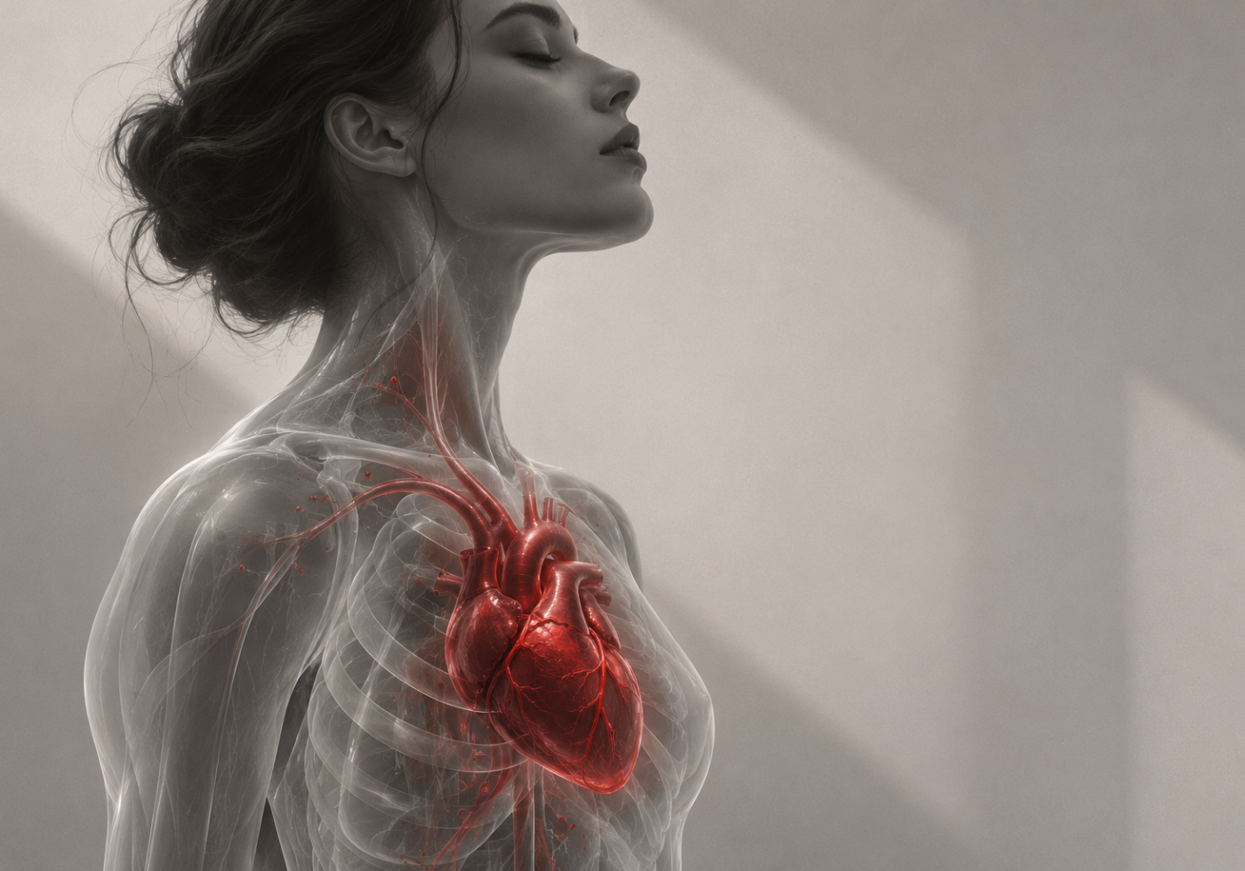
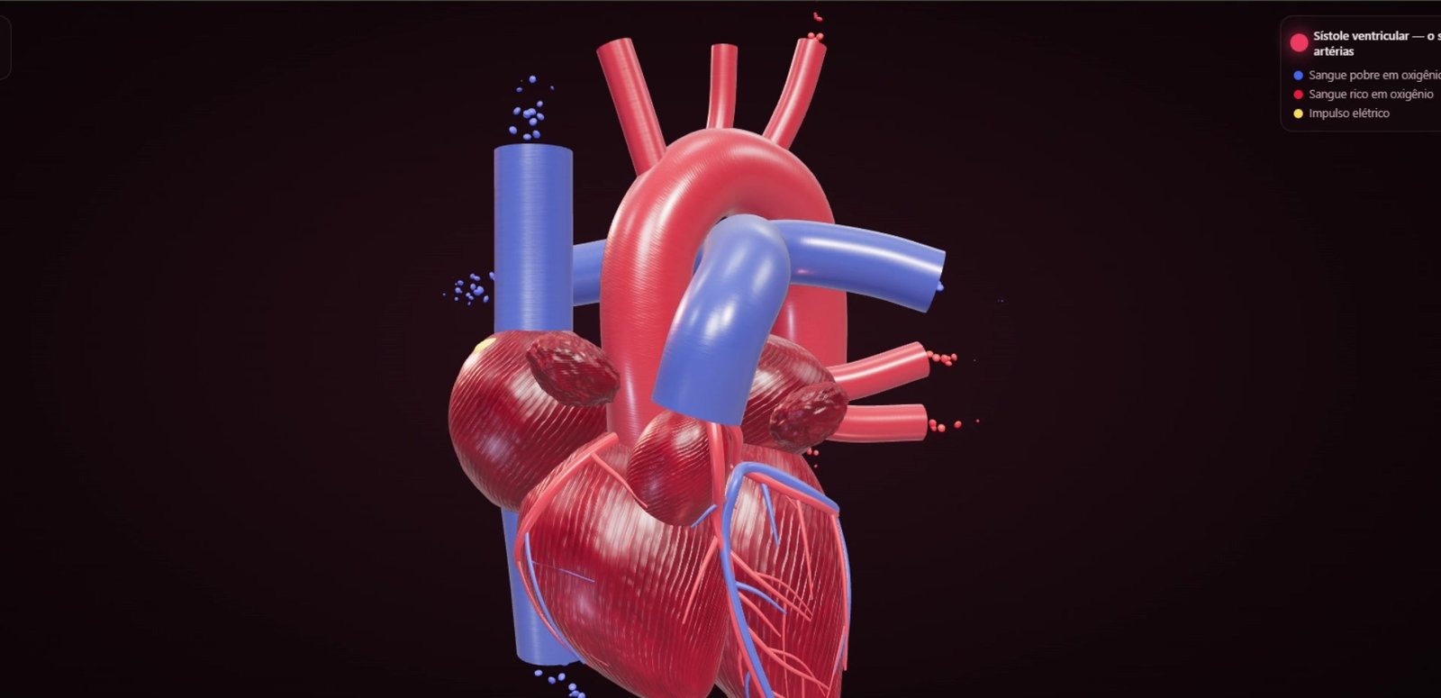
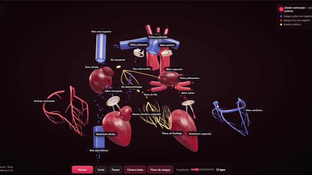
A working single-file WebGL experience with 21 labelled anatomical structures, four chambers, four valves, cardiac conduction, adjustable heart rate and 1,300 animated blood-flow particles.
This independent experimental project explores how browser-based 3D and AI-assisted development can turn cardiac anatomy into an explorable visual system. Instead of loading an external 3D model, the heart is constructed procedurally from code and rendered in real time in the browser.
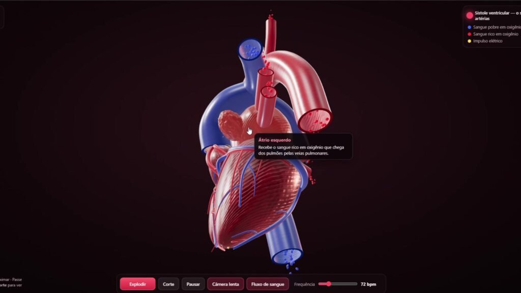
The result combines anatomical structures, cardiac motion, blood circulation and electrical conduction in one interactive experience. Visitors can rotate and zoom the model, inspect labelled structures, change the simulated heart rate and switch between assembled, internal and exploded views.
What visitors can explore
Rotate and zoom the heart directly in the browser.
Hover over or touch structures to see their names and functions.
Explode and reassemble the model to examine its internal organisation.
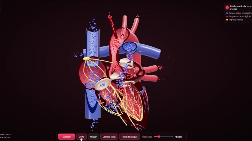
Activate a cutaway view to reveal internal chambers, valves and circulation.
Pause the animation or view it in slow motion.
Adjust the simulated heart rate from 40 to 180 beats per minute.
Toggle the animated blood flow.
Anatomical system
The experience contains 21 labelled anatomical structures. These include the four cardiac chambers, four valves, the aorta, pulmonary trunk, venae cavae, pulmonary veins, coronary arteries, cardiac veins and the main components of the cardiac conduction system.
The conduction sequence represents the sinoatrial node, internodal pathways, atrioventricular node, bundle of His, right and left bundle branches and Purkinje fibres. Animated red and blue particles distinguish oxygen-rich and oxygen-poor blood as it moves through the model.
Technical approach
The complete experience is contained in a single HTML document of approximately 1,200 lines. It uses Three.js and WebGL for real-time rendering, physically based materials for the surfaces, clipping planes for the internal cutaway, CSS2DRenderer for anatomical labels and GLSL-based effects for the electrical pulse.
The blood-flow system uses 1,300 instanced particles distributed across the circulation paths. Tissue colour, texture and surface relief are generated procedurally with mathematical noise rather than image textures. No external 3D model was used.
AI-assisted development
Artificial intelligence supported the ideation, coding and iterative development process. The work still required defining the visual direction, structuring the anatomy, testing interactions, identifying failures and refining the final experience as a coherent browser-based project.
Outcome
The final result is a responsive WebGL prototype that brings together anatomical visualisation, motion design, interaction design and front-end development. It demonstrates how code and AI-assisted workflows can be combined to build an original 3D experience without specialist modelling software or pre-built anatomical assets.
This project is an educational visualisation and creative technology experiment. It is not a medically validated anatomical model and must not be used for diagnosis, clinical decision-making or formal medical instruction without specialist review.